——Faster, More Precise, More Reliable
Image Registration and Electrode Calibration
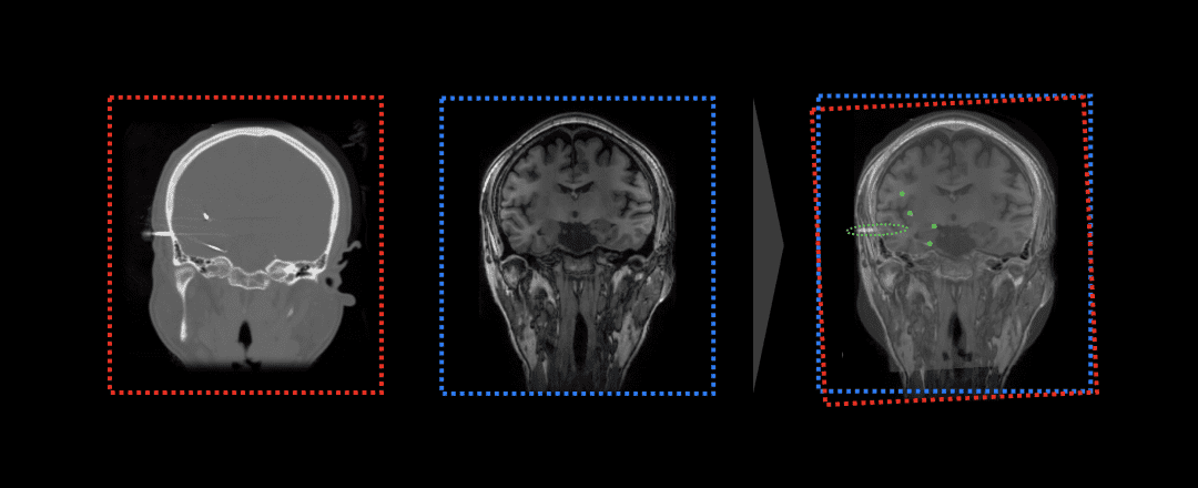
Registering MRI and CT images before and after SEEG implantation surgery, deeply aligning cranial structures in different modal images, making electrode contacts in CT images visible in the intracranial soft tissue distribution of MRI. The registration process requires fine feature extraction, feature matching, coordinate transformation, and optimized interpolation in different modal images to ensure registration accuracy. The EEG-X intelligent EEG analysis platform provides adaptive rigid and elastic registration functions. Simply upload the patient's pre-implantation MR examination results and post-operative CT images to select the appropriate solution with one click and complete the data registration operation, obtaining the intracranial soft tissue distribution map of electrode contacts.


Calibrating electrodes in CT images after SEEG electrode implantation, combined with the transformation matrix obtained from registration, can map electrode contact positions to intracranial soft tissue distribution, providing precise location information for epilepsy monitoring, lesion diagnosis, and subsequent intervention treatment. Traditionally, SEEG electrode calibration is mainly completed through manual fine operation, requiring doctors to find and align electrode contacts in CT slice images of different views one by one, which is labor-intensive, tedious, has high training and learning costs, and is prone to mislabeling and missing labels. The EEG-X intelligent EEG analysis platform, leveraging CT image 3D reconstruction technology and powerful statistical analysis algorithms, fully exploits the spatial distribution information, morphological information, and prior information of SEEG electrode images, achieving adaptive electrode filtering, reshaping, and segmentation, and possessing full-automatic, high-precision, and strong-flexibility calibration capabilities for electrode contacts. Doctors can obtain complete, accurate, and comprehensive electrode contact coordinate sequences with one click using EEG-X, greatly saving clinical work time and effort.
When calibrating electrodes with unclear contact contours, EEG-X has higher resolution compared to manual calibration. Based on 3D point cloud image reconstruction, the algorithm can detect tiny features in the data with powerful statistical analysis technology, sensitively capturing electrode contact positions, as precise as "lidar", surpassing the limits of human visual perception. Additionally, EEG-X's algorithm's adaptive coordinate correction technology can perform region-adaptive correction on found contacts, enabling the algorithm to accommodate cases where implanted electrodes are curved in actual situations, eliminating systematic coordinate errors and greatly improving the algorithm's robustness and practical value.
