Non-Invasive Source Localization

Rapid Source Localization Based on Interictal Spikes

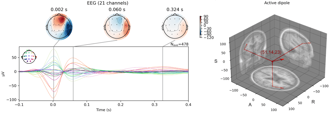
EEG source localization is a non-invasive method that uses scalp-recorded potential signals to inversely calculate the location, direction, and intensity of intracranial neural activity sources. In clinical research, EEG source localization has become an important tool for preoperative assessment of drug-resistant epilepsy patients due to its non-invasive nature and ease of operation, and has proven its value in guiding epileptogenic zone localization and SEEG implantation planning in multiple studies. In practice, it commonly analyzes interictal spike discharge events identified through non-invasive methods such as electroencephalography (EEG) and magnetoencephalography (MEG) to provide estimates of the irritative zone. This technology also plays a key role in localizing high-frequency oscillations, motor imagery, working memory, and other brain functional area activity studies.
Non-Invasive Source Localization
Multiple Source Localization Methods Integration - More Precise, More Convenient

EEG-X integrates multiple EEG source localization methods, including traditional MNE, sLORETA, and the patented DeESI algorithm based on statistical bias correction. Compared to traditional algorithms, DeESI provides superior source localization accuracy and temporal signal estimation. Users can freely switch between localization methods based on their preferences and data characteristics, with one-click preview of suspicious intracranial discharge locations. The algorithm includes two standard adult brain templates, enabling rapid analysis using standard templates even with EEG data only. The highly integrated source localization module performs fully automated analysis throughout the process, requiring no additional operations.

Multiple Source Localization Methods Integration - More Precise, More Convenient
Digital Twin of Whole Brain Structure

High-level preoperative assessment requires precise modeling of patient-specific data to achieve personalized surgical planning. EEG-X integrates multimodal three-dimensional head model analysis solutions, capable of tissue segmentation using T1-weighted magnetic resonance images, reconstructing three-dimensional conduction fields, and providing high-precision forward model solutions for EEG source localization, thereby improving the quality of source lesion localization. If patients also have T2-weighted magnetic resonance images or CT scan data, EEG-X provides fully automatic image fusion registration solutions, utilizing rich imaging information to enhance source localization model accuracy.